How Did the Prophet ﷺ Know? Embryology, Genetics, and the Hadith of the Nutfah
The Prophet ﷺ described, fourteen centuries ago, biological realities that Western science only confirmed in the twentieth century — among them the precise mechanism of fetal sex determination, the six-week threshold of gonadal differentiation, and the indestructibility of the primitive streak (عجب الذنب), the very structure that modern embryology identifies as the organizer and builder of the human body.
This research paper was compiled by Dr. Ahmad al-Shami (22/4/2023) and draws exclusively on peer-reviewed studies, authoritative embryology textbooks, and primary scientific literature.
Table of Contents
- Part One — Fetal Sex Determination (جنس الجنين)
- Definition of Sex Determination
- Factors Affecting Sex Determination
- The Sex Reversal Phenomenon
- The Role of SRY
- Gonadal Differentiation Does Not Begin Until After the Sixth Week
- First Production of Estradiol and Testosterone — Seventh Week
- The Role of WNT4
- The Role of SOX9
- Anti-Müllerian Hormone (AMH)
- Insulin-like Hormone-3 (INSL3)
- Then We Created of the Clot Another Thing
- Part Two — The Bones (العظام)
- Part Three — The Eye (العين)
- Part Four — The Role of the Ovum in Sex Determination
- Part Five — ‘Ajab al-Dhanab — The Primitive Streak
Part One — Fetal Sex Determination (جنس الجنين)
Definition of Sex Determination
Sex determination is a cascading molecular process, not a single event — it begins at fertilization but its visible effects do not manifest until the sixth week of embryonic life.
Peter Goodfellow and Robin Lovell-Badge, among the most celebrated geneticists of the twentieth century — famous for discovering the role of the SRY gene in sex determination — define it as follows:
“Sex determination is the process of choosing either the male or the female sexual differentiation pathway.”
— ADVANCES IN GENOME BIOLOGY: Volume 4. GENETICS OF SEX DETERMINATION, by RAM S. VERMA, The Long Island College Hospital–SUNY Health Science Center Brooklyn, New York, VIII. DEVELOPMENTAL PROCESSES: SEX DETERMINATION AND DIFFERENTIATION, pg13.
The eminent embryologist Benjamin Willier offers the following definition:
“Zygotic determination of sex is meant the primary determining constitution established in the fertilized egg by the union of the gametes…; that is the nuclear determiners or genes which decide whether male or female sex characters shall develop…. By sexual differentiation is meant the origin or expression of sex differences during ontogeny.”
— Willier, B.H. The embryonic development of sex. In: Sex and Internal Secretions (Allen, E., Ed.), 2nd ed., Baillière, Tindall and Cox, London, 1939, pp.13–14.
The following diagram from Developmental Biology (6th edition) illustrates the complete gonadal differentiation pathway — from the undifferentiated gonadal ridge through to either testis or ovary, governed by a cascade of genes including SRY, SOX9, DAX1, WNT4, and SF1:

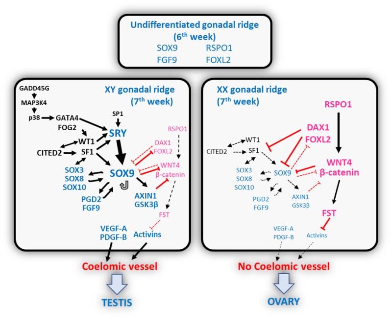
The second diagram shows the full genetic pathway in both the XY (7th week) and XX (7th week) gonadal ridge, with all regulatory genes mapped:
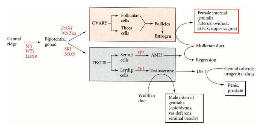
Source: Developmental Biology, 6th edition — Chromosomal Sex Determination in Mammals
Factors Affecting Sex Determination
The process of sex determination in mammals operates through three distinct, sequential stages:
“The process of sex determination in mammals normally unfolds in three distinct stages: (1) establishment of chromosomal sex at fertilization (XX or XY); (2) commitment to the appropriate pathway of gonadal differentiation with respect to chromosomal sex, through the action (or absence) of the Y chromosome gene SRY; and (3) correct development of secondary sexual characteristics, including internal and external genitalia, in accordance with gonadal sex. At any of these three steps, the process of sex determination can go awry, leading to disorders of sexual development.”
— Koopman P., abstract. The molecular genetics of sex determination and sex reversal in mammals. By Quinn A. europepmc.org
The three factors are:
-
Chromosomal sex
-
Gonadal sex
-
Development of external and internal genitalia
The Sex Reversal Phenomenon
XX males and XY females are a documented biological reality in mammals including humans — a finding that demonstrates sex determination is a genetic process requiring active molecular instruction, not a passive consequence of chromosome possession.
“The fortuitous findings of XX males and XY females, which are generally termed sex reversal phenomenon, are quite bewildering traits that have caused much amazement concerning the pairing mechanism(s) of the pseudoautosomal regions of human X and Y chromosomes at meiosis.”
— ADVANCES IN GENOME BIOLOGY: Volume 4. GENETICS OF SEX DETERMINATION, by RAM S. VERMA, The Long Island College Hospital–SUNY Health Science Center Brooklyn, New York, intro, pg XIII
“Many examples of XX males and XY females are known in different mammalian species, including the human one.”
— ADVANCES IN GENOME BIOLOGY, Vol. 4, VII. SEX REVERSAL, pg10–11
Regarding the Dosage-Sensitive Sex Reversal Gene (DSS):
“One of these causes failure of testis development when present in double dose on the same X chromosome. It has been named DSS (dosage sensitive sex reversal gene).”
— ADVANCES IN GENOME BIOLOGY, Vol. 4, XI. TESTIS-DETERMINING GENES, pg18
The Role of SRY
SRY expression commences between days 41 and 44 post-fertilization in humans — a fact corroborated by no fewer than five independent research teams across two decades.
“The SRY gene product, SRY, is detected in the bipotential gonad of XY individuals at about 42 days.”
— Hormones, by Anthony W. Norman and Helen L. Henry, pg267. Google Books
“The chromosomal sex of the embryo is established at fertilization. However, 6 weeks elapse in humans before the first signs of sex differentiation are noticed. Sex differentiation involves a series of events whereby the sexually indifferent gonads and genitalia progressively acquire male or female characteristics. Believed initially to be governed entirely by the presence or absence of the SRY gene on the Y chromosome…. The presence or absence of primordial germ cells, of extragonadal origin, also has a sexually dimorphic relevance. Subsequently, internal and external genitalia will follow the male pathway in the presence of androgens and anti-Müllerian hormone (AMH), or the female pathway in their absence.”
— Sexual Differentiation, by Rodolfo Rey, MD, PhD, Nathalie Josso, MD, PhD, and Chrystèle Racine, PhD., abstract. ncbi.nlm.nih.gov/books/NBK279001/
“Owing to its Y-chromosome localization, SRY can only be expressed in the XY gonadal ridge, thus playing a paramount role in tilting the balance between testicular and ovarian promoting genes towards the male pathway. A tight regulation of SRY expression is essential for fetal gonadogenesis: both timing and level of expression are determinant, as revealed by experiments in mouse showing that SRY levels must reach a certain threshold at a certain stage of fetal development to induce testis differentiation. SRY expression commences between days 41 and 44 post-fertilization in humans.”
— Sexual Differentiation — Sex-Determining Region on the Y Chromosome (SRY). ncbi.nlm.nih.gov/books/NBK279001/
This information is corroborated by a dedicated study from the University of Manchester and Newcastle University by Dr. Neil Hanley, Dr. Donna Hagan, and ten additional researchers, entitled “SRY, SOX9, and DAX1 expression patterns during human sex determination and gonadal development, 2000”:
“SRY expression commences in the gonadal ridge of 46,XY embryos between 41 and 44 d.p.o./CS17–18. Peak SRY expression is detected at 44 d.p.o./CS18, when sex cords are first visible, thereby defining testicular determination.”
— Hanley NA, Hagan DM, Clement-Jones M, Ball SG, Strachan T, Salas-Cortes L, McElreavey K, Lindsay S, Robson S, Bullen P, Ostrer H, Wilson DI. SRY, SOX9, and DAX1 expression patterns during human sex determination and gonadal development. Mech. Dev. 2000;91:403–407. sciencedirect.com
The following diagram from that study summarizes the role of SRY and shows how the state of testicular and ovarian formation changes before and after the end of the sixth week:
A 1990 study published in Letters to Nature by 13 researchers stated:
“In conclusion, a de novo mutation in the gene SRY is associated with sex reversal in an XY female. This provides compelling evidence that SRY is required for testis formation and male sex determination.”
From a study entitled “A human XY female with a frameshift mutation in the candidate testis-determining gene SRY” (1990):
“The primary decision about male or female sexual development of the human embryo depends on the presence of the Y chromosome, more specifically on a gene on the Y chromosome encoding a testis-determining factor, TDF. ….In this region there is a candidate gene for TDF, termed SRY, which is conserved and specific to the Y chromosome in all mammals tested….We have now identified a mutation in SRY in one out of 12 sex-inversed XY females with gonadal dysgenesis who do not lack large segments of the short arm of the Y chromosome.”
From a study by Robin Lovell-Badge and Elizabeth Robertson on “XY female mice resulting from a heritable mutation in the primary testis-determining gene, Tdf”:
“In mammals, it is known that the activity of a gene on the Y chromosome is responsible for determining the primary sex of the developing embryo (Jacobs and Strong, 1959; Ford et al. 1959; Welshons and Russell, 1959).”
“In the absence of the Y chromosome, the bipotential gonad follows the ‘default’ ovarian pathway, which has been postulated to involve a set of ovary-determining genes (Eicher and Washburn, 1986)…. There are also many cases of human XY females. Some of these are due to chromosomal abnormalities; for example, where the region carrying TDF has been lost due to abnormal X:Y interchange at meiosis. Others may represent mutations within TDF.”
“SRY is found in normal XY males and in the rare XX males, and it is absent from normal XX females and from many XY females.”
“SRY encodes a high-motility group (HMG) box type protein that controls the expression of other genes including the steroidogenic factor 1 (SF1), Wilms’ tumor-related gene (WT1), HMG-box 3 (SOX3) and HMG-box 9 (SOX9) required for testicular development.”
Gonadal Differentiation Does Not Begin Until After the Sixth Week
Male and female embryos are morphologically indistinguishable until Day 42 — confirmed by every major obstetrics and embryology textbook.
“Until approximately 42 days, Carnegie stage 17, this ridge forms the indifferent gonad, and male and female embryos are morphologically indistinguishable.”
“The gonad remains undifferentiated until around 42 days.”
“Male and female embryos are morphologically indistinguishable till 42 days.”
“The appearance of the primitive gonad is similar in both sexes until 42 days after fertilization.”
“No sexual difference can be observed in the gonads until the 6th week of embryonic life in humans. Undifferentiated gonads of XX or XY individuals are apparently identical and can form either ovaries or testes. This period is therefore called the indifferent or bipotential stage of gonadal development.”
“In the 6th week of embryonic life, the gonadal ridge is sexually undifferentiated, and various factors are expressed at the same levels in the XX and the XY gonads. During the 7th week, in the XY gonad, SRY expression is triggered, and the male pathway prevails…. In the XX gonad, the female pathway prevails.”
“Sexual differentiation begins at ~6 weeks post conception (p.c.) (Witschi, 1963; Wartenberg, 1982; Lovell-Badge and Robertson, 1990), governed by the sex-determining region on the Y-chromosome (Lovell-Badge and Robertson, 1990; Koopman et al., 1990).”
“In the female gonad the indifferent period terminates between day 40 and 42 of ovulation age (20 to 23 mm CR length). Between day 40 and 50 the blastemal content of the indifferent gonad is remodelled and an ovarian cortex differentiates.”
The same study provides the following table of prespermatogonia counts beginning from Week 6:
| Fetal age p.c. (weeks + days) | Fetal ID | No. of prespermatogonia | No. of Sertoli cells |
|---|---|---|---|
| 6 + 0 | 1 | 2,890 | — |
| 7 + 0 | 2 | 13,459 | 133,712 |
| 7 + 4 | 3 | 15,510 | 178,372 |
| 8 + 0 | 4 | 25,708 | 395,771 |
| 8 + 1 | 5 | 36,340 | 338,122 |
| 8 + 2 | 6 | 25,593 | 287,313 |
| 8 + 2 | 7 | 19,677 | — |
| 8 + 2 | 8 | 26,338 | — |
| 9 + 0 | 9 | 17,498 | — |
| 9 + 1 | 10 | 40,585 | 431,645 |
“Irrespective of their sex-chromosome constitution, all mammalian embryos are anatomically, histologically and functionally undifferentiated from a sexual standpoint during the first stages of development, approximately six weeks in humans and 10 days in mice.”
First Production of Estradiol and Testosterone — Seventh Week
Functional hormonal differentiation begins at the seventh week — even before histological differentiation is visible in the XX gonad.
“In the XX fetus, the gonad remains histologically undifferentiated after the 7th week from a histological standpoint, but a functional differentiation is already detectable: XX gonads become capable of estradiol production at the same time as XY gonads begin to synthesize testosterone.”
“Leydig cells are first found at about 60 days of gestation. Leydig cells secrete testosterone, the regulator of male differentiation.”
“In Leydig cells, SF1 activates the expression of steroidogenic enzyme systems from the 8th week of gestation, which results in androgenization of the external genitalia.”
“Secretion of testosterone by the interstitial Leydig cells begins at approximately 9 weeks of gestation.”
“Androgen secretion by the fetal testis is essential for male phenotypic differentiation. In the human fetus testosterone formation is initiated soon after the differentiation of the testis (approximately 8 weeks of gestation).”
See also: Textbook of Medical Physiology, 3rd Edition, by Indu Khurana, Arushi Khurana, Narayan Gurukripa Kowlgi, pg729. Google Books
The Role of WNT4
“The absence of WNT4 in female embryos (XX) leads to masculinization.”
— Cloning and characterization of wnt4a gene and evidence for positive selection in half-smooth tongue sole (Cynoglossus semilaevis). nature.com/articles/srep07167
Individuals who have a duplication of the chromosome containing the wnt4 gene exhibit sex reversal.
The Role of SOX9
“Gain-of-function of SOX9 in XX individuals leads to sex reversal.”
“On the other hand, increased expression of SOX9 in XX gonads induces testicular differentiation resulting in foetal virilisation even in the absence of SRY…. Male differentiation can occur in XX individuals in the presence or the absence of SRY.”
“XX humans who have an extra copy of SOX9 develop as males, even though they have no SRY gene (Huang et al. 1999).”
“6–7 WEEKS: SRY expression and SOX9 expression transforming progenitor cells to pre-sertoli cells (testes determination). Also role of (WT1, SF1, DMRT1/2, DHH, ATRX, SOX8, ARX). 7 weeks: Sertoli cell differentiation (Fgf9 & Fgfr2) leads to primary sex cords, phenotypic sex.”
“In mammals, the pre-Sertoli cell of the male genital ridge is the first cell type to display sex-specific differentiation and differential gene expression.”
— Presumptive pre-Sertoli cells express genes involved in cell proliferation and cell signalling during a critical window in early testis differentiation.
Anti-Müllerian Hormone (AMH)
“Anti-Müllerian hormone (AMH) is a glycoprotein hormone secreted by pre-Sertoli cells of the testis from around the seventh week onwards.”
“Sertoli cells….from the 7th week onwards, these secrete anti-Müllerian hormone.”
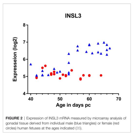
Insulin-like Hormone-3 (INSL3)
“This gene may be involved in the development of urogenital tract and female fertility.”
— National Library of Medicine, National Center for Biotechnology Information. ncbi.nlm.nih.gov/gene/3640
The following graph shows INSL3 mRNA expression levels measured by microarray analysis of gonadal tissue from individual male (blue triangles) and female (red circles) human fetuses across fetal age in days post-conception:
“By week 8 of gestation, two key supporting cells of the testis can be identified: Sertoli cells, which line the seminiferous tubules, and Leydig cells, which populate the interstitium. Two key gene products are secreted from these cells that direct the future function of the gonad and the final determination of the internal genital ductal system. The first hormone, produced by Sertoli cells, is mullerian inhibitory substance….. Sertoli cells also express desert hedgehog gene, a molecule that plays an important role in spermatogenesis. Secretion of testosterone by the interstitial Leydig cells begins at approximately 9 weeks of gestation.”
Then We Created of the Clot Another Thing
“When the embryo becomes 7 or 8 weeks old, it is known as a ‘foetus’.”
Overexpression of DMRT1 in XX mice inhibits WNT4 and FOXL2 expression and results in partial testicular differentiation and male genital development. (Sexual Differentiation, by Rodolfo Rey, Nathalie Josso, Chrystèle Racine, abstract.)
The following image shows the embryo at Carnegie Stage 10 (approximately the 7–8 week mudgha/foetus transition), sourced from the Endowment for Human Development:
Reference: ehd.org/developmental-stages/stage10.php
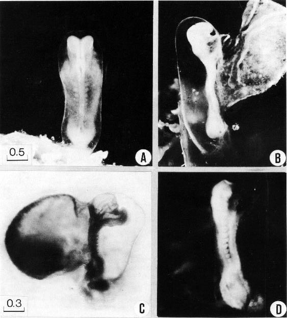
The following two panels show human embryo developmental stages (A, B, C, D) from the clinical embryology atlas:
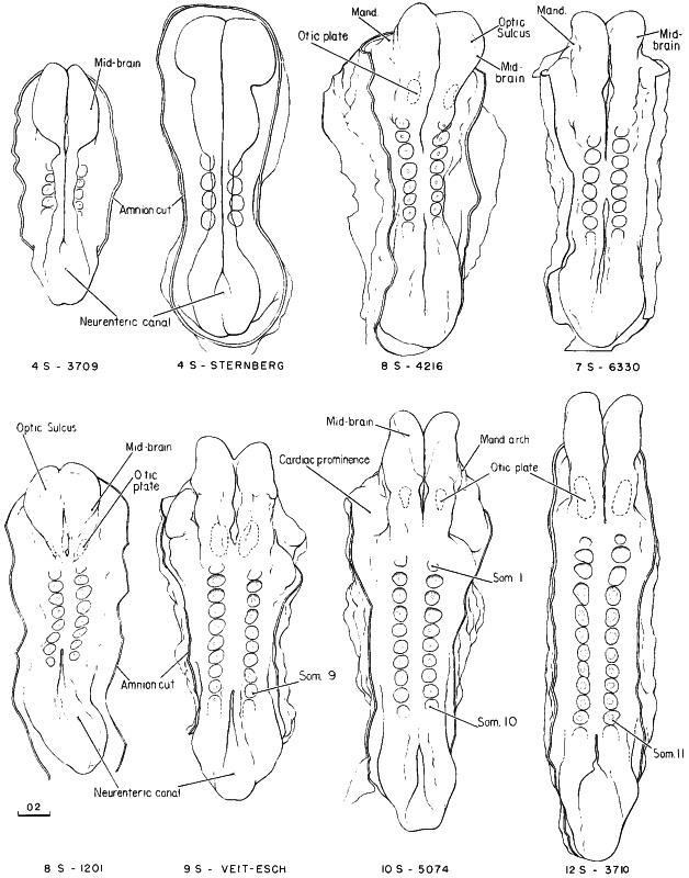
The Hamburger-Hamilton stage comparison below shows corresponding developmental stages across 4S–12S in the chick embryo model, used in comparative embryological research:
Reference: researchgate.net — Hamburger-Hamilton stage 10 chick embryo showing development of somites
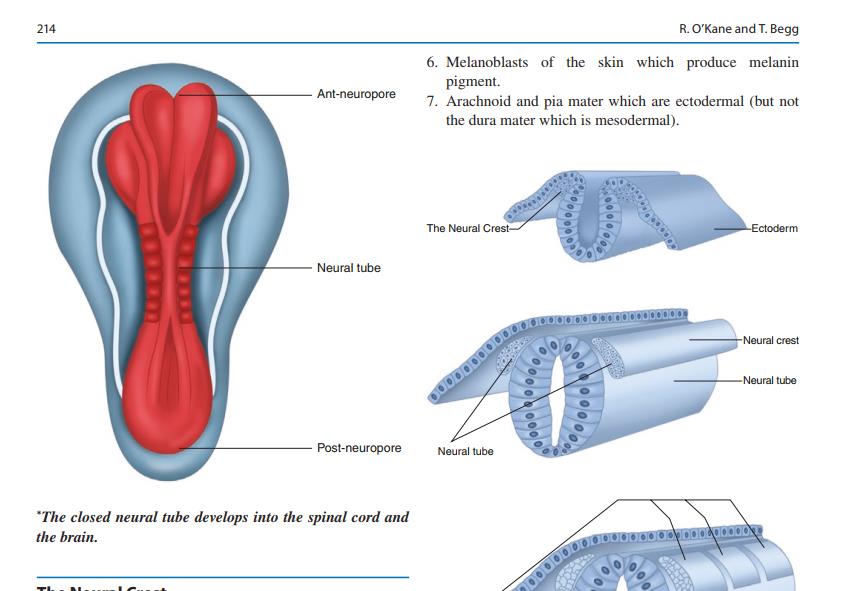
Part Two — The Bones (العظام)
(A complete detailed separate study is available)
The Clavicle — First Bone to Ossify
The clavicle is the first bone in the human skeleton to begin ossification — appearing at approximately Day 39, at the end of the sixth week of intra-uterine life.
“Weeks 6–7: The clavicle (first bone to ossify) begins to ossify.”
“The clavicle is probably the first bone in the human skeleton to show evidence of bone formation, which can be detected in the differentiating mesenchyme from approximately day 39 in the sixth week of intra-uterine life.”
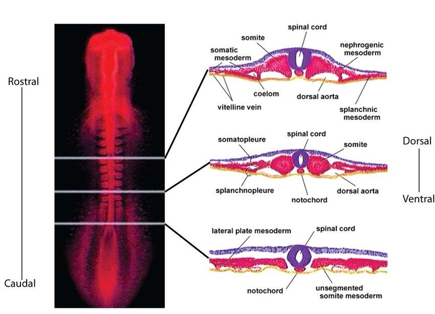
The following diagram from Clinical Embryology An Atlas of Congenital Malformations shows the neural tube development and related bone formation anatomy:
Part Three — The Eye (العين)
The duration of eye formation extends from approximately the first week to the seventh week.
The Eight Parts of the Eye
Eye Anatomy: Parts of the Eye and How We See; By Kierstan Boyd, David Turbert; Reviewed By Ninel Z Gregori, MD — aao.org
The diagram below identifies the eight anatomical components of the human eye — all of which begin development by the seventh week:
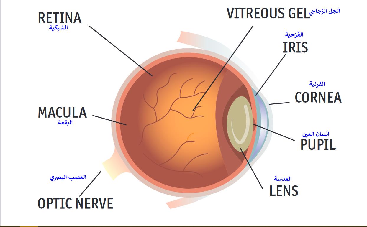
“At about 7 weeks, the main parts of the eye that enable sight – the cornea, iris, pupil, lens, and retina – start developing.”
— How your baby’s eyes and vision develop in the womb. babycenter.com
Stages of Eye Formation — Week by Week
The three major phases of eye development are:
-
Embryogenesis — formation of primary organ rudiments, ending at the end of Week 3
-
Organogenesis — development of primary organ rudiments, extending to the end of Week 8
-
Differentiation — division of each primitive organ into a fully or partially functional organ
“The first period called embryogenesis is characterized by the establishment of the primary organ rudiments and ends at the end of the 3rd week with the appearance of the optic sulci on both sides of the midline at the expanded cranial end of the still open neural folds. The second period called organogenesis includes the development of the primary organ rudiments and extends till the end of the 8th week. The third period involves the differentiation of each of the primitive organs into a fully or partially active organ and is called differentiation.”
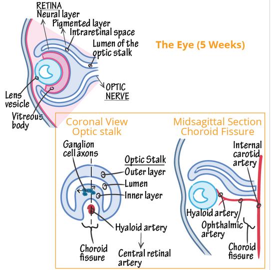
The cross-sectional diagram below shows the eye at Week 5, with the optic stalk, lens vesicle, vitreous body, retinal and choroid fissure all visible:
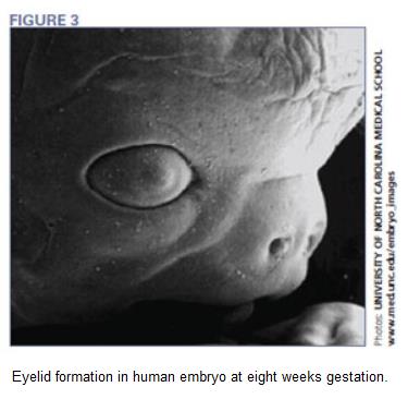
The image below shows eyelid formation in a human embryo at eight weeks gestation:
“The human eye begins to develop during the 17th day of gestation.”
— Embryonic Eye Development. 2020mag.com
“Eye development occurs in the human embryo from approximately the third week through the tenth week of gestation.”
“The appearance of optic grooves (neuroectoderm origin) from the developing forebrain marks the first sign of eye development at week three of gestation.”
Week 2 — Formation of the Eyeball Begins:
“The eyeball development begins early in the 4th week of intrauterine life with formation of optic vesicle (a diverticulum) from diencephalon.”
“Embryologic eye development begins at 4–5 weeks of gestation (27–36 days) during lens vesicle formation.”
“To all that activity, add the formation of the orbits and extraocular muscles at four weeks gestation.”
“Early in the 4th week optic vesicles extend from the 3rd ventricle and wall of the forebrain (diencephalon). As the vesicle reaches the surface ectoderm it flattens (a) and progressively invaginates (b) to form the optic cup.”
The following information panel records the key events at Day 25:

Week 3 — Trilaminar Disc, Optic Sulcus, Retinal Pigmentation:
“By the third week of development, the three germ layers are arranged in a flat disc-like structure called the trilaminar disc, which marks a key milestone towards the formation of the developing embryo.”
“Pigmentation of the retinal epithelium occurs at approximately week 3 or 4; this is the earliest pigmentation evident in the embryo.”
The Day 33 panel below records the closure of the embryonic fissure — a key developmental milestone:

Week 4 — Eyelids Begin, Lens Placode (Day 27), Choriocapillaris:
“At approximately week six of gestation the eyelids begin to form.”
“The choriocapillaris starts to differentiate simultaneously with the development of the retinal pigment epithelium during the fourth and fifth week.”
Week 5 — Lacrimal Glands, Lens (Day 35), Sclera, Hyaloid Artery, Neuroblastic Layers:
“The lens begins development from an out-pouching of the surface ectoderm at approximately day 35.”
“At seven weeks, the sclera develops from embryonic tissue.”
“A branch of the internal carotid artery enters the optic cup through the fetal fissure to become the hyaloid artery during week 5.”
“At approximately week 7, cell migration occurs, forming the inner and outer neuroblastic layers, between which lies the transient fiber layer of Chievitz.”
Week 6 — Optic Stalk to Brain (Day 36), Lacrimal Glands, Ganglion Cell Axons, Optic Nerve:
“The optic stalk, the precursor to the optic nerve, forms a critical connection to the forebrain at 36 days.”
“Formation of optic nerve occurs between sixth and eighth weeks.”
“8 weeks: Tear ducts start to develop.”
“By week 8, a second wave of mesenchyme proliferates, migrates between the developing epithelium and endothelium, and gives rise to the fibroblasts, collagen, and ground substance of the stroma.”
“Although rarely seen, pericytes can be detected as early as the sixth week.”
The comprehensive eyelid development timeline table below, compiled from 11 sources, maps all eyelid structural events from Week 6 through Week 20:
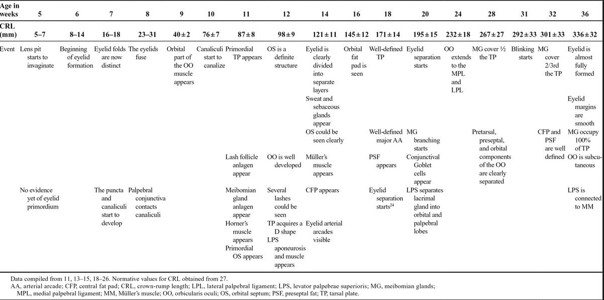
Reference: ncbi.nlm.nih.gov/pmc/articles/PMC5102278/
Week 7 — Completion of Iris, Anterior Tunica Vasculosa Lentis, Basal Lamina:
“Between 30 and 35 days, you can see the start of the iris. In two more weeks, it is fully developed.”
“The annular vessel sends loops forward onto the anterior surface of the lens to form the anterior vascular tunic of the lens (anterior tunica vasculosa lentis) during the seventh week.”
“The first component of the anchoring system is the basal lamina, which is evident by week 9, and hemidesmosomes are present by week 13.”
Week 10 — Bowman’s Layer, Iris Epithelium, Pupillary Membrane:
“At 3 months all layers of the cornea are present, except Bowman’s layer, which appears during the fourth month.”
“By the end of the third month, the lip of the optic cup begins to elongate and grows between the lens and the developing cornea. The outer layer of the optic cup becomes the anterior iris epithelium and the inner layer forms the posterior iris epithelium.”
“During the third month, the pupillary membrane forms between the lens epithelium and the corneal endothelium to replace the vascular tunic.”
Week 11 — Fusion of Eyelid Folds:

Week 14 — Zonular Fibers, Lamina Cribrosa, Aqueous Humor, Trabecular Meshwork:
“During the fourth month, vessels of hyaloid system atrophy progressively. Zonular fibers (tertiary vitreous) begin to stretch from the growing ciliary region toward the lens capsule.”
“During the fourth month connective tissue fibers cross the posterior scleral foramen, running through the optic nerve fibers and producing the first connective tissue strands of the lamina cribrosa.”
“Aqueous humor production begins at 4 to 6 months of gestation.”
“The trabecular meshwork is visible as a triangular mass of mesenchymal cells during the fourth month.”
Week 18 — Ciliary Muscle, Myelination:
“The ciliary muscle begins to develop from neural crest during the fifth month.”
“Myelination of the axon begins during the fifth month of gestation once the fiber reaches the lateral geniculate nucleus.”
Week 20 — Eyelid Separation, Photoreceptor Cells:
“During the fifth month, a two-month process begins — the separation of the eyelids.”
“The photoreceptor cells are the last cells of neural retina to differentiate; this occurs during the fifth month.”
Part Four — The Role of the Ovum in Sex Determination
The Ovum Selects the Sperm
The egg is not a passive recipient — it actively selects sperm based on genetic content — a finding that challenges the foundational assumption of Mendelian random gamete union.
“The oldest law of genetics says that gametes combine randomly, but experiments hint that sometimes eggs select sperm…”
— Choosy Eggs May Pick Sperm for Their Genes, Defying Mendel’s Law. quantamagazine.org
This is based on research by Dr. Joseph H. Nadeau of Oxford University, published under the title “Do Gametes Woo? Evidence for Their Non-random Union at Fertilization”:
“His hypothesis — that the egg could woo sperm with specific genes and vice versa — is part of a growing realization in biology that the egg is not the submissive, docile cell that scientists long thought it was. Instead, researchers now see the egg as an equal and active player in reproduction.”
“These unusual results suggest that fertilization is genetically biased toward particular gametes based on their genetic content.”
— Do Gametes Woo? Evidence for Their Nonrandom Union at Fertilization. academic.oup.com
“The female reproductive tract has the capacity to select and orient sperm, making it an active recipient of male gametes.”
— The Control of Male Fertility by Spermatozoon Ion Channels. ncbi.nlm.nih.gov/pmc/articles/PMC3914660/
Vaginal pH and Fetal Sex
Normal vaginal pH range: 3.8–4.5
A study entitled “Effect of Vaginal pH in Preconceptional Fetal Sex Determination” by Dr. Mohammed Gaber (Professor of Dermatology and Venereology, Menoufia University) and Dr. Saeed Salah (Professor of Obstetrics and Gynecology, same faculty) concluded that fetal sex can be determined based on vaginal pH. Study parameters:
-
20 patients from Menoufia University clinics, ages 21–37 years
-
Excluded: women with conditions affecting vaginal pH (diabetes, vaginitis, cervicitis, other significant medical problems)
-
Complete obstetric, gynecological, and medication history taken
-
Study group: 20 women at ovulation time
-
Vaginal pH measured using litmus paper at first days of ovulation
-
Vaginal ultrasound used to measure ovarian follicle diameter (reaching 20mm at ovulation) along with luteinizing hormone measurement
-
Women divided into alkaline and acidic vaginal pH groups
-
After determining fetal sex by ultrasound, correlation with acidic and alkaline vaginal pH was established
The results table below shows a statistically significant association (p = 0.007) between fetal male sex and alkaline vaginal pH, and fetal female sex and acidic vaginal pH:
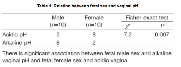
Reference: mmj.eg.net — Effect of Vaginal pH in Preconceptional Fetal Sex Determination
Differences Between X and Y Sperm
X-bearing spermatozoa are statistically larger, longer, and wider than Y-bearing spermatozoa.
“Statistically, the length, perimeter and area of the sperm heads, and the length of the sperm necks and tails of X-bearing spermatozoa were significantly larger and longer than those of Y-bearing spermatozoa.”
This finding was confirmed by Joep P.M. Geraedts, Professor of Genetics and Molecular Biology at Maastricht University, Netherlands, in a study entitled “X spermatozoa larger than Y in 1973”:
X spermatozoa larger than Y in 1973.
Part Five — ‘Ajab al-Dhanab — The Primitive Streak
The primitive streak is the most consequential structure in early human embryogenesis — it organizes the entire body plan, determines all cell fates, and is identified by modern science as the iconic structure from which every tissue and organ of the human body originates.
The following photograph is among the earliest real images of the primitive streak:
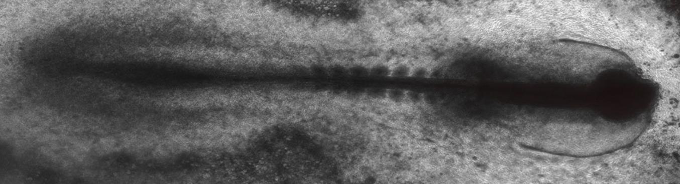
Reference: discovery.lifemapsc.com — Embryonic Development of the Primitive Streak
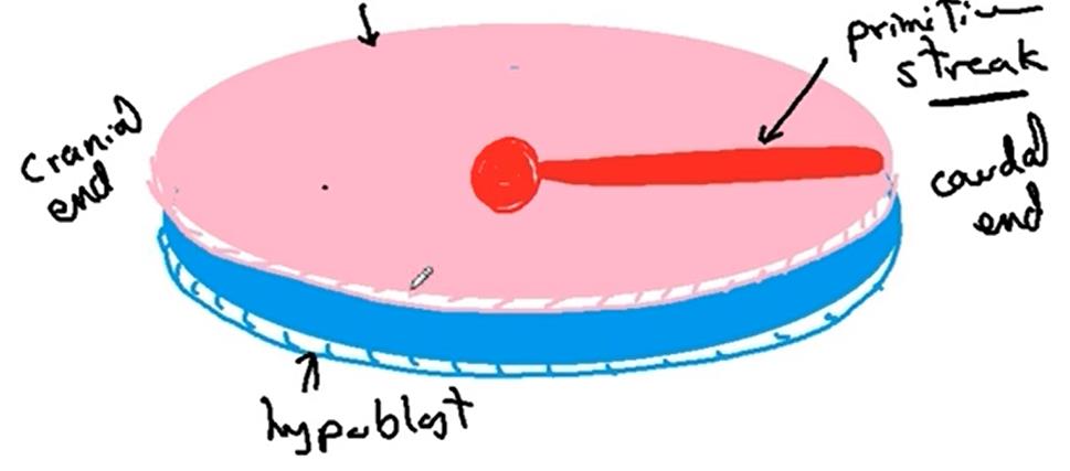
The diagram below illustrates how every structure of the body arises from the primitive streak, situated at the caudal end of the hypoblast disc:
Functions of the Primitive Streak
The primitive streak is:
-
The builder of all body cell types — it forms every cell type of the human body
-
The marker of human individuality — its appearance at Day 14 is recognized internationally as the beginning of individual human identity
-
The legal threshold in the 14-day rule — many nations establish Day 14 as the limit for experimental intervention with human embryos, as it marks the appearance of the primitive streak
-
The controller of cell fates — it determines which cell becomes which tissue and organ
-
The builder of the germinal layers — it produces the three germ layers (endoderm, mesoderm, ectoderm) from which all cells arise
-
The source of developmental commands — it issues the molecular instructions required for brain formation and organ differentiation
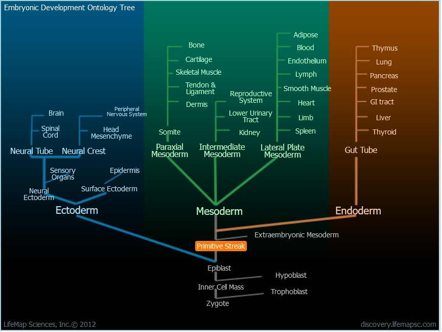
The following Embryonic Development Ontology Tree shows how every structure of the body — from bone, blood, brain, lung, kidney, heart, and liver to the nervous system, epidermis, and reproductive system — traces back through the primitive streak to the zygote:
“The primitive streak also gives anterior-posterior and dorsal-ventral spatial information to cells undergoing gastrulation and forming the various body cell types…symbolize the emergence of human individuality. As such, many countries have established a legal limit of 14 days for the in vitro culture of fertilized human eggs — this is known as the ‘14-day rule.’”
The study describes it as an iconic structure.
— The primitive streak and cellular principles of building an amniote body through gastrulation. science.org/doi/10.1126/science.abg1727
“After fertilization, a human embryo implants into the uterus (day 6–12) and gastrulation starts soon afterwards, with the primitive streak emerging at about day 14. Gastrulation allocates cell fates … laying down the foundation of the human body.”
“One of the most critical periods of development…occurs at approximately two weeks after fertilization through a process called gastrulation and is associated with an embryonic structure called the primitive streak — a structure in early development that initiates bilateral symmetry and germ layer formation.”
“Gastrulation is defined as an early developmental process in which an embryo transforms from a one-dimensional layer of epithelial cells (blastula) and reorganizes into a multilayered and multidimensional structure called the gastrula…gastrulation derives a three tissue-layered organism composed of endoderm, mesoderm, and ectoderm…is marked by the appearance of a groove in the caudal end of the epiblast layer known as the primitive streak.”
In 2018, researcher Kelly Servick wrote in Science magazine an article entitled “Elusive Master Organizer of Human Embryo Growth” — science.org — in which she considered the primitive streak as the answer to the question posed by Brivanlou:
“‘Where does the information come from … to make a brain or discrete organs?’ Brivanlou says.”
— Elusive master organizer of human embryo growth seen for the first time. 23 May 2018. science.org
“The embryo forms a furrow called the primitive streak and folds inward on itself while cells mature into different lineages that will later give rise to all the organs and tissues of the body.”
— merriam-webster.com
“The primitive streak … defines the future embryonic midline and serves as a conduit of cell migration for germ layer formation…. Once the initial primitive streak is established, germ layer formation begins.”
“Gastrulation is a process of cellular rearrangement which involves migration, invagination and differentiation of the epiblast. It is largely controlled and orchestrated by the primitive streak. The primitive streak is a groove in the midline of the epiblast which appears during the third week. Within the primitive streak lies a primitive node at the cranial end, and within the primitive node lies the primitive pit. Cells of the epiblast layer break off and migrate toward the primitive pit. Here, they detach and penetrate through the epiblast layer to form three new germ cell layers: Endoderm — formed by epiblast cells that migrate through the primitive pit and displace the hypoblast cells. Mesoderm — formed by epiblast cells that migrate through the primitive pit and lie between the epiblast layer and the newly created endoderm. Ectoderm — formed by the epiblast cells that remain in position. These three cell layers are then responsible for forming the different tissues of the fetus.”
— teachmeanatomy.info
Study of Spemann and Mangold
Study title: “Induction of Embryonic Primordia by Implantation of Organizers from a Different Species” by Hans Spemann and Hilde Mangold.
Reference: ijdb.ehu.es/article/11291841
Every Son of Adam Will Decay Except the Coccyx
The primitive streak retains its inductive capacity even after being killed by heat, crushing, or coagulation — a fact established through multiple independent experiments.
The Prophet ﷺ said: “Every son of Adam will decay except the coccyx — from it he was created, and from it he will be reconstructed.”
C.H. Waddington — embryologist, geneticist, and founder of systems biology — in his article “Experiments on Embryonic Induction” recorded:
“Bautzmann, Holtfreter, Spemann and Mangold (1932) had shown that in the Amphibia coagulated organisers are still able to exert the inductive capacities which they possess in the living state. This work followed on the earlier experiments of Marx (1931) who obtained inductions with narcotised organisers, and Spemann (1931a) who found that the inductive capacity is retained when the organiser tissue is subjected to crushing.”
— Experiments on Embryonic Induction: Part II. Experiments on Coagulated Organisers in the Chick. journals.biologists.com
Waddington himself conducted two experiments by killing the organizer with heat and found it still retained its inductive capacity:
“The two successful experiments 32–331 and 33–17 described above are sufficient to show that in the chick the capacity to perform an induction can be present in dead material.”
— Experiments on Embryonic Induction: Part II. Experiments on Coagulated Organisers in the Chick. journals.biologists.com
This proves that the cells of the coccyx (primitive streak / عجب الذنب) were not destroyed by these processes. What was destroyed in the experiments: spinal cord cells, fat cells, and muscle cells — while the primitive streak’s inductive core remained active.
This research paper will continue to be updated, God willing.
Dr. Ahmad al-Shami — 22/4/2023