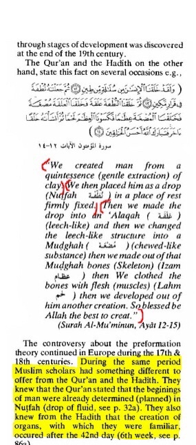
Surah Muminun 14 Embryology
Here we go:
Facts are, in embryology, the very first bones to form are the vertebrae, a major part of the skull and ribs.
All of these appear prior to any sort of striated muscle tissue appearing. Especially since they largely do not
form the same way other bones do - the processes are different, which means other factors of synthesis are applied
(such as intermembraneous ossification, which does not require an elongated step-by-step process,
like the endochondral ossification does).
So, it is a fact the rib cage, a large part of the skull and the vertebrae form before any striated muscle tissue,
again. What about the limbs?
The limbs start to elongate by the proliferation (multiplication) of mesenchymal cells. These mesenchymal cells
very quickly differentiate into chondrocytes. Chondrocytes inherently make up hyaline cartilage, which we can
already accept as bone (since it’s cartilage).
After the differentiation of chondrocytes, specific somites differentiate and migrate to the parts of the limbs
where chondrocytes are already forming cartilage. There, they take a multi-step process before becoming muscle cells -
but that’s not muscle tissue (flesh).
In fact, the muscle cells become flesh only when they form the actually tissue - when myofibrils are formed.
By the formation of myofibrils, we can be sure that the mesenchymal mass in the center of the limb is already
cartilage.
SUMMARY: A part of the skull, the ribs and the vertebrae form before any striated muscle tissue.
In the limbs, cartilagenous axis is established first, and only after differentiated somites migrate
into these same parts of the limb, slowly encapsulating (wrapping around) the cartilage by forming myofibrils
(striated muscle tissue).
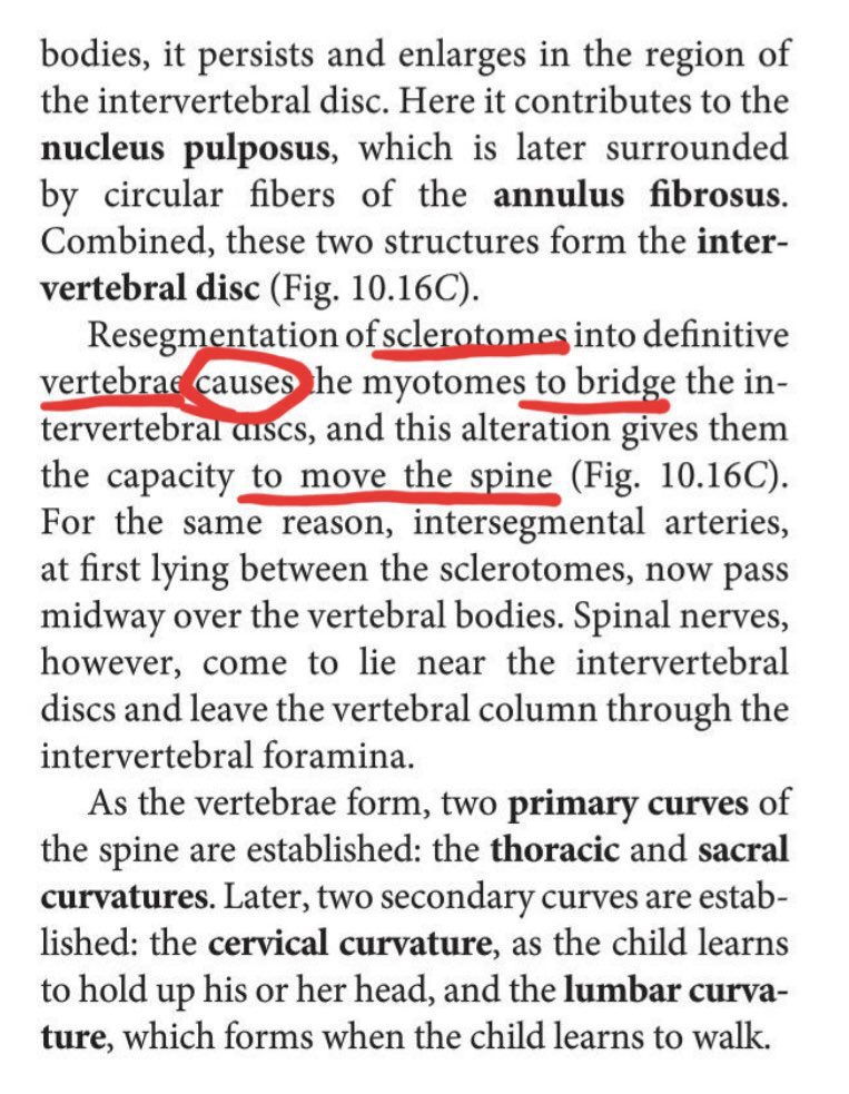
This illustration clearly implies that the sclerotome forms before the dermomyotome develops,
and way before the dermomyotome further develops (separates) into the dermatome and myotome layers.
In fact, it clearly shows how large and developed the sclerotome is while the mesenchyme cells on the outer
rim aren’t even yet differentiated.
Of course, there are different types of the differentiation of somatomes and whatnot,
but I wouldn’t go too specific.
(Scleratome = bone layer
Dermatome = skin layer
Myotome = muscle layer)
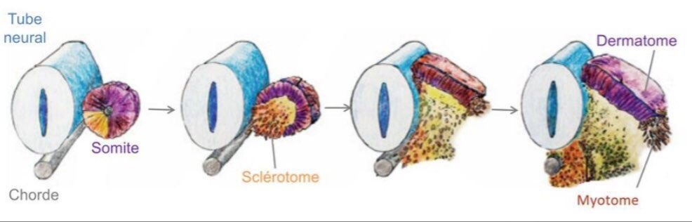
Another illustration, which is portrayed a little differently, but actually entails the same information.
The red part is the paraxial mesoderm (undifferentiated cells - mesenchymal cells), which we can clearly later
see mentioned as the dermatome (skin layer).
The second bit in the image already contains a differentiated mesoderm centre with chondrocytes (cartilagenous axis)
and an undifferentiated, not-yet-even-developed dermomyotome.
The dermomyotome is mentioned in the third bit, as that’s when it develops. Again, we can see a very large
scleratome layer already formed (cartilagenous axis).
Finally, in the third bit is the myotome finally separated and recognised as differentiated. Way after the
cartilagenous axis is already established.
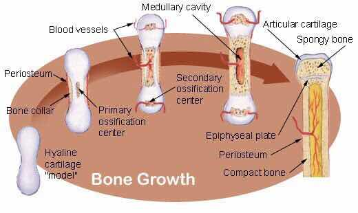
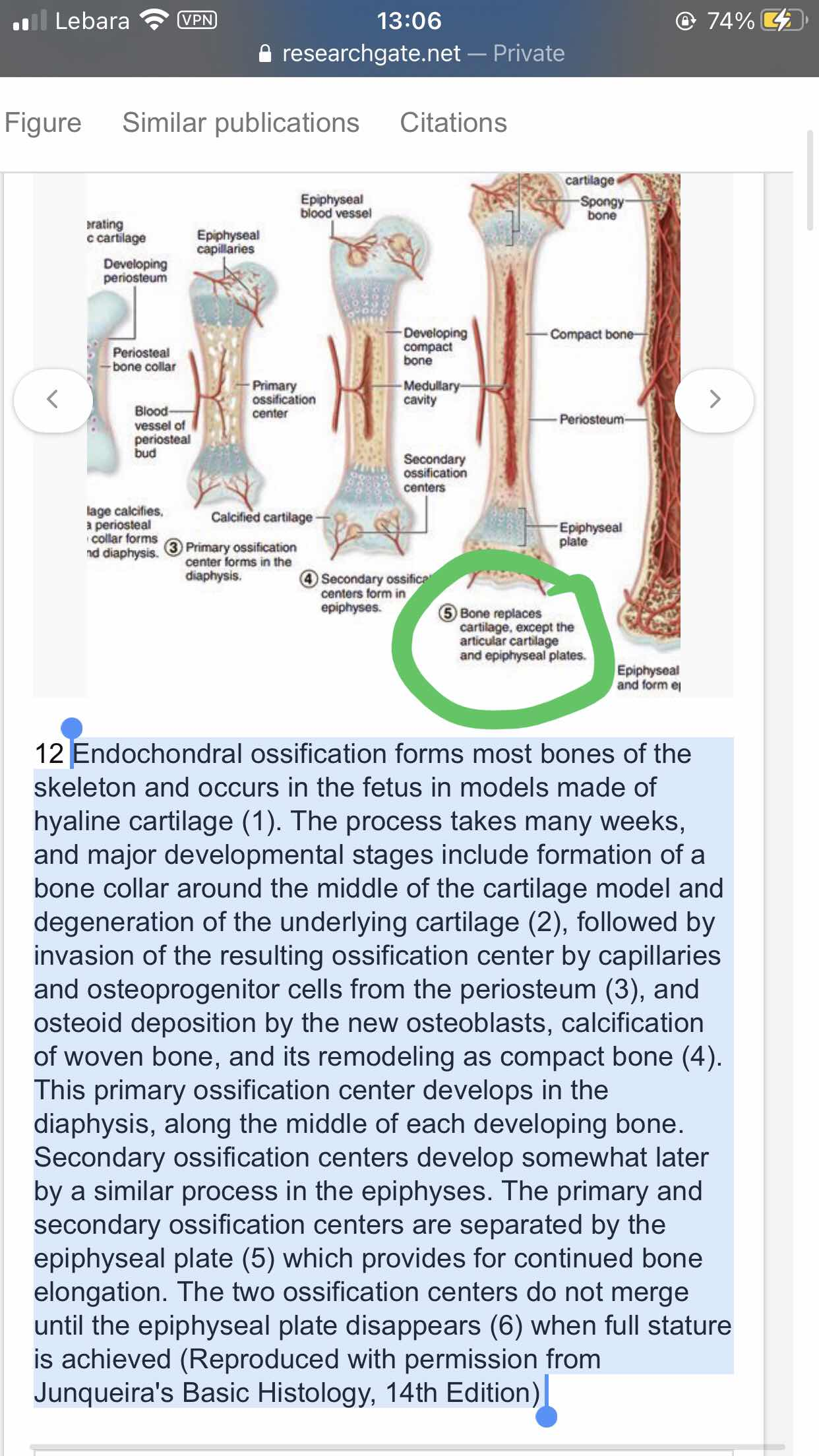
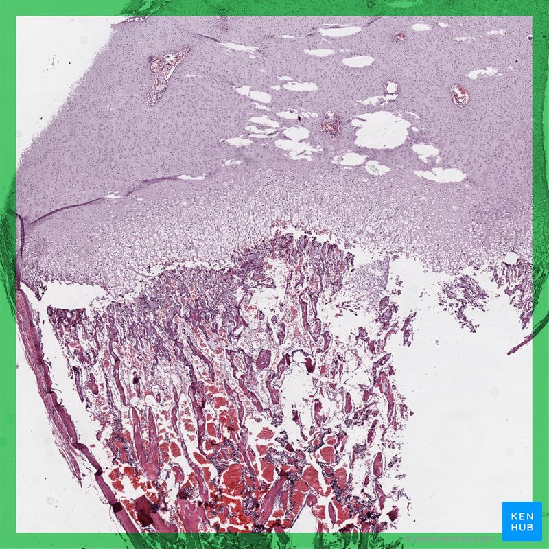
An image depicting the process of the formation of the bone, more specifically, the differentiation of the mesenchyme
cells into chondrocytes and after, those further developing into ossified bone.

An image depicting certain stages of limb development.
As I’ve previously stated - the loose mesenchyme forms the outside part of the limb bud and elongates the limb
while condensed mesenchyme forms inside it by gene activation.
From this condensed mesenchyme, mostly chondrocytes (cartilagenous tissue) forms. Meanwhile, the somites which
differentiate into myoblasts undergo a procedure of, well, differentiation and migration into the limb buds.
Only after arriving at the limb buds, the myoblasts start to synthesise in order to create further muscle tissue.
This is also why most “limb development” images focus on the development of the bone structure and have zero emphasis on muscle.
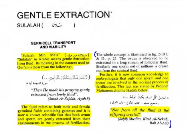
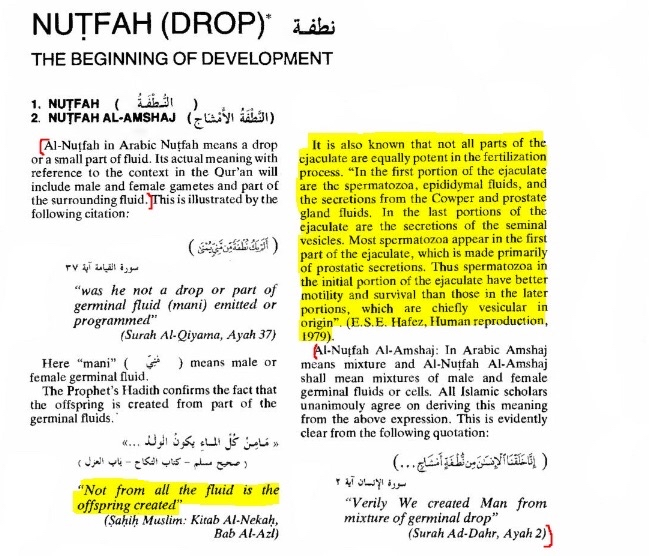
The Qur’ān doesn’t mention sperm metazoan, the word nutfah which is most of the time translated into sperm drop, even the Arabs
in their text books when the they talk about sperm they call it “al-Haywan al-Manawi ” because nuftah
is not specific enough to be the egg which we know of and see in the diagrams, now the reason is imagine
of the people at the time read that there’s small animals in your sperm, they would go crazy and ask
“what the hell ”
This is simply evidence that God was aware of how much the Arabs knew and understood,
so he used simple words so that they may understand. Side Note: Whats very interesting is that the sperm
and the ovam and the egg are all contained in the fluid.
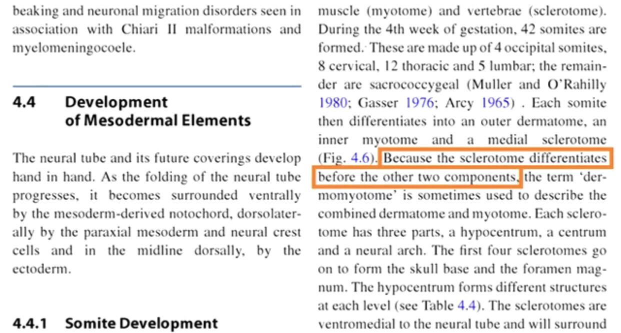
In the developing vertebrate embryo, somites split to form dermatomes, skeletal muscle (myotomes),
tendons and cartilage (syndetomes) and bone (sclerotomes). Because the sclerotome differentiates before
the dermatome and the myotome, the term dermomyotome refers to the combined dermatome and myotome before
they too separate out
https://emedicine.medscape.com/article/1287982-overview
{Embed}
https://emedicine.medscape.com/article/1287982-overview
Hand Embryology: Gross Morphologic Overview of Upper Limb Developme…
Classic descriptions of upper limb development and embryology relied solely on detailed descriptions of the gross morphology of the developing embryo. A developmental process is a morphologically observable event such as limb bud formation or the development of digits.
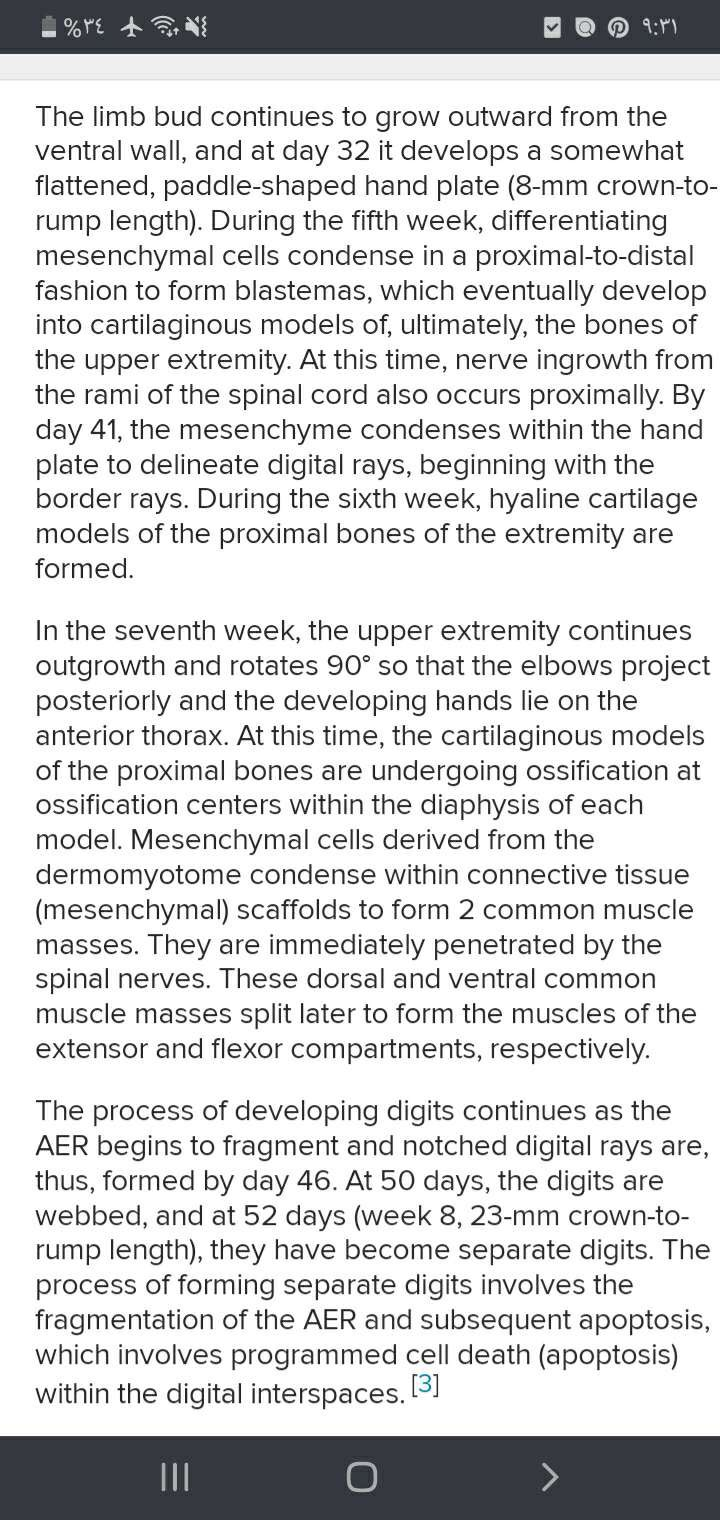
Mesoderm within the developing limb bud
differentiates initially to form cartilage which
later ossifies by endochondral ossification. Hypaxial somitic mesoderm from somites at the levels of limb
bud formation, migrates into the bud. These cells within the bud proliferate in regions of muscle formation,
fuse to form myotubes and then differentiate to form
skeletal muscle cells.
The mesoderm was NOT bony at first. It was flesh like, and so it is mentioned. The skeletal
muscle cells are differentiated later on, not simultaneously with the differentiation
of cartilage.
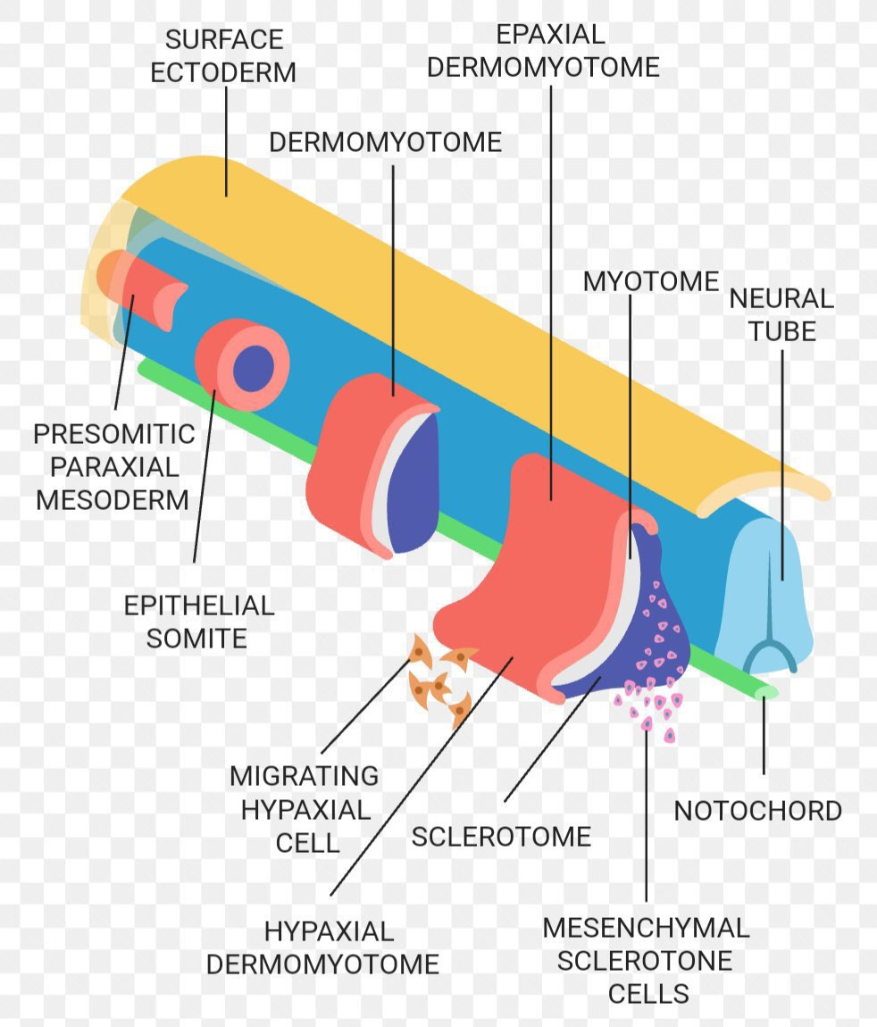
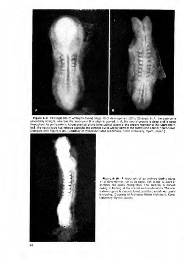
Somites appear bilaterally as pairs at the
same time and form earliest at the cranial
(rostral,brain) end of the neural groove and add sequentially at the caudal end. This
addition occurs so regularly that embryos are staged according to the number of somites
that are present. Different regions of the
somite differentiate into dermomyotome (dermal and muscle component) and sclerotome (forms vertebral column).
An example of a specialized musculoskeletal
structure can be seen in the development of the limbs.musculoskeletal and limb abnormalities are on of the
largest groups of congenital abnormalities
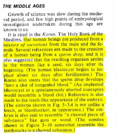
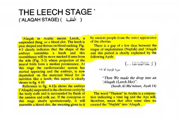
Now for the alaquah claim the Qūr’ān is describing how it looks not that it is literally a clot and clots look
like leaches and that is correct
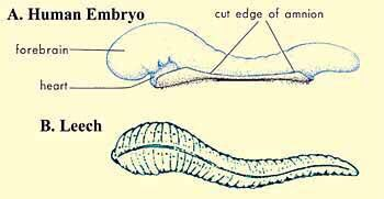
So either it is alaqah that is the leech or clot both are correct
https://archive.org/details/developing-human-islamic/
The%20Developing%20Human%20%283rd%20ed.%2C%201983%29%3B%20With%20Islamic%20Additions%20%28Keith%20L.
%20Moore%3B%20Abdul-Majeed%20A.%20Azzindani%29%20%28low-size%3B%20OCR%27d%29/
{Embed}
https://archive.org/details/developing-human-islamic/
The Developing Human; With Islamic Additions (Keith L. Moore; Abdul…
Keith L. Moore and Abdul-Majeed A. Azzindani, The Developing Human: Clinically Oriented Embryology; With Islamic Additions: Correralation Studies With Qur’an…

Read the unhighlighted parts asw the hyaline cartilage will eventually either remain as it is such as with
Meckel’s cartilage or replaced by bone
https://pubmed.ncbi.nlm.nih.gov/3315379/
{Embed}
https://pubmed.ncbi.nlm.nih.gov/3315379/
Earliest evidence of cartilage and bone development in embryonic li…
Some aspects of the development of cartilage and bone during embryonic life are discussed in this review and an attempt is made to show that studies of development, even when performed on species far removed from humans, are relevant to clinical orthopedic surgery. Initially, some definitions of ske …

https://www.ncbi.nlm.nih.gov/books/NBK539718/
{Embed}
https://www.ncbi.nlm.nih.gov/books/NBK539718/
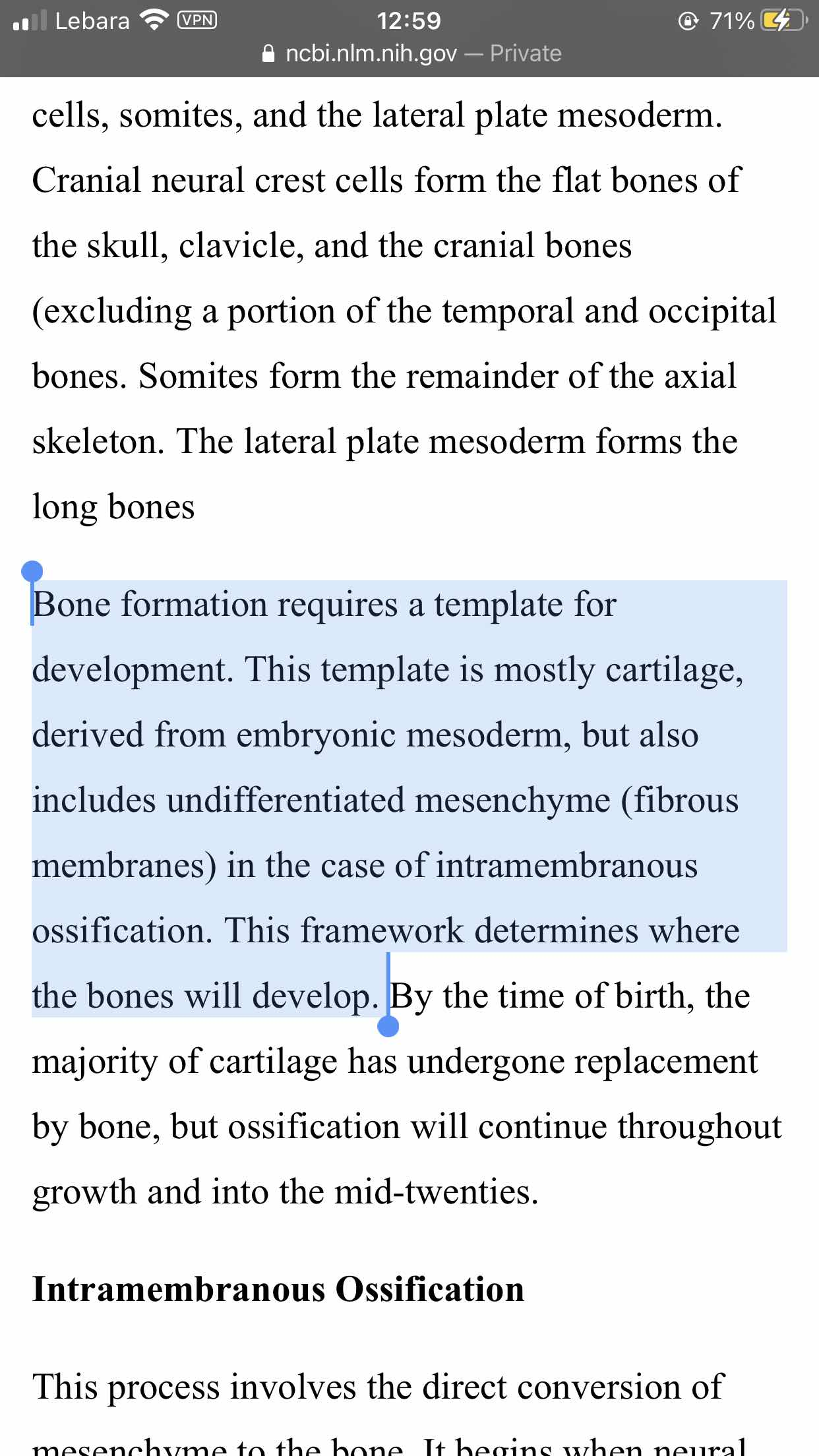
Embryology, Bone Ossification
Bone ossification, or osteogenesis, is the process of bone formation. This process begins between the sixth and seventh weeks of embryonic development and continues until about age twenty-five, although this varies slightly based on the individual. There are two types of bone ossification: intramembranous and endochondral. Each of these processe…

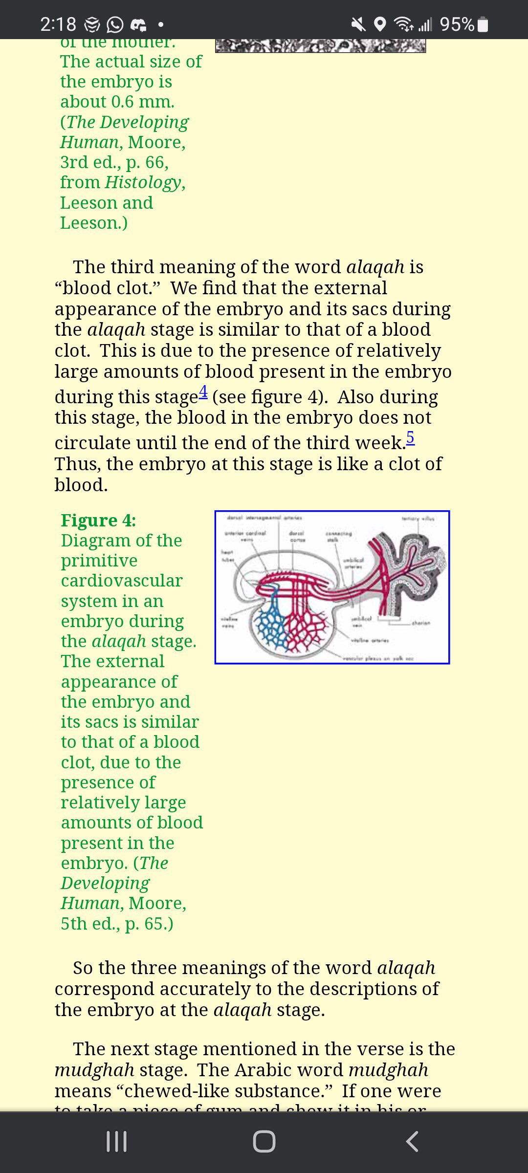
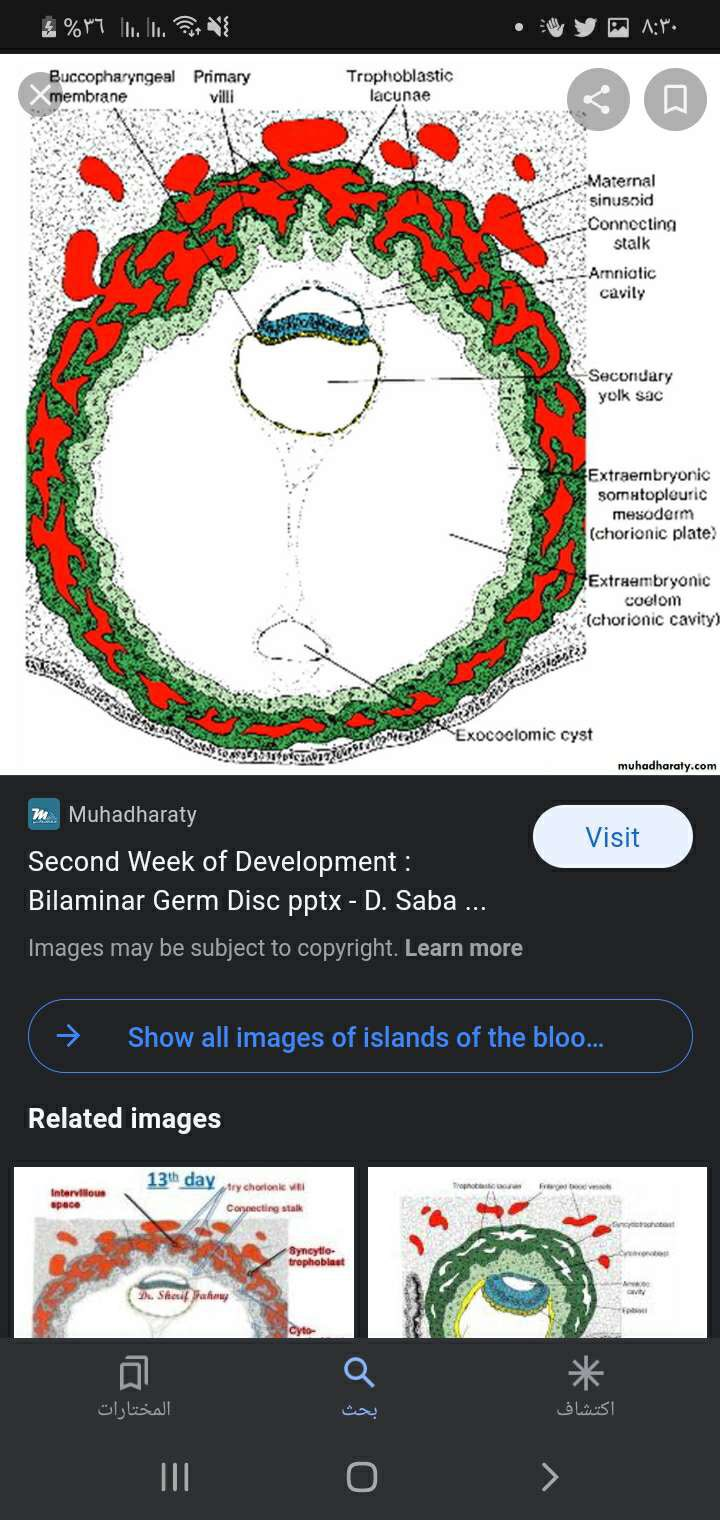
The first verse says that he made a mixture between the sperm and the egg a clut like structure called ( alaka )
Scientifically this example is very accurate because between day 13 to 15 we absorb type formation of what
we call {blood islands}
Blood islands are clusters of cells that arise from the mesoderm layer of the developing embryo and are the
precursors to blood vessels and blood cells.
The blood islands form in the yolk sac, which is one of the extra-embryonic membranes that surrounds the
developing embryo. The yolk sac is responsible for producing blood cells during the early stages of embryonic
development until the liver and bone marrow take over this function later in development
{Embed}
Extraembryonic Blood Islands - Cellular Development, Function & Ana…
Learn about Extraembryonic Blood Islands development @ Lifemap Discoveryanatomical and cellular development of the Extraembryonic Blood Islands
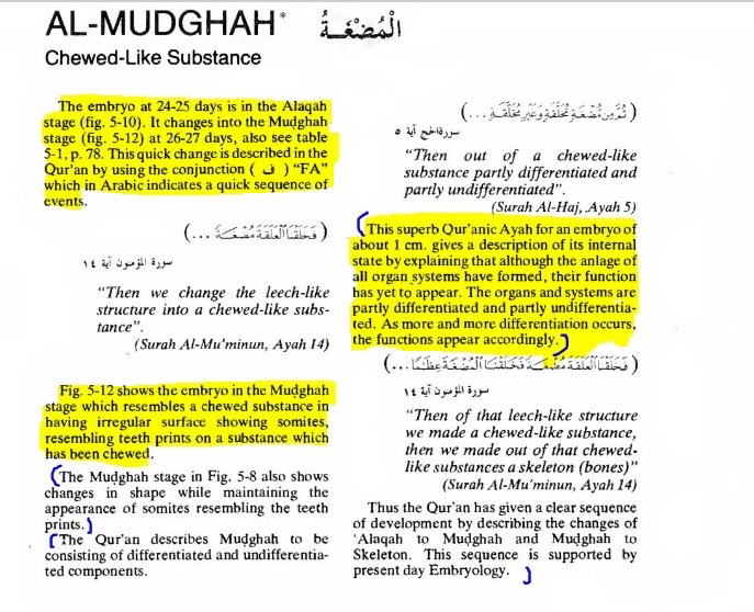
The second verse says then he made the alaka (mudga) which is structure of different tissues with no specific
shape at this stage the emperionic structure will form the heart at the day 20 which cause the the circulatory
system to work and allows the clustered blood at the stage of blood Islands to move and circulate so it will
move toward the next stage from a lot like structure to a structure who has meat of muscle of the heart and
circulating blood instead of clustered blood next to other tissues
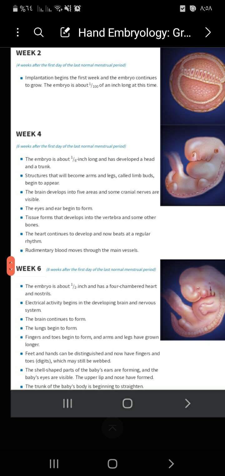
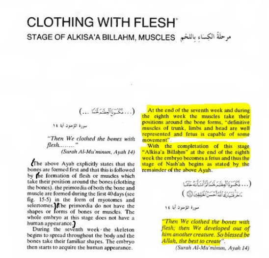
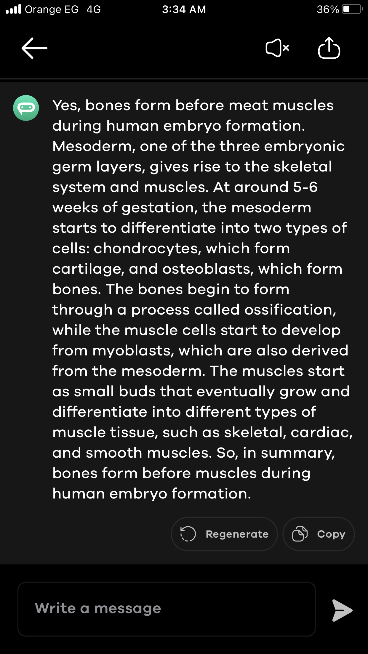
And do the final claim Quran makes the claim that bones are formed before muscles by saying that he made bones
in the alaka them covered these bones by muscles which is scientifically true because bones are firstly formed
at the first half of the embryonic structure between week 6 to 7 well skeletal muscles are just formed between
week 10 to 13
https://pubmed.ncbi.nlm.nih.gov/23622355/
{Embed}
https://pubmed.ncbi.nlm.nih.gov/23622355/
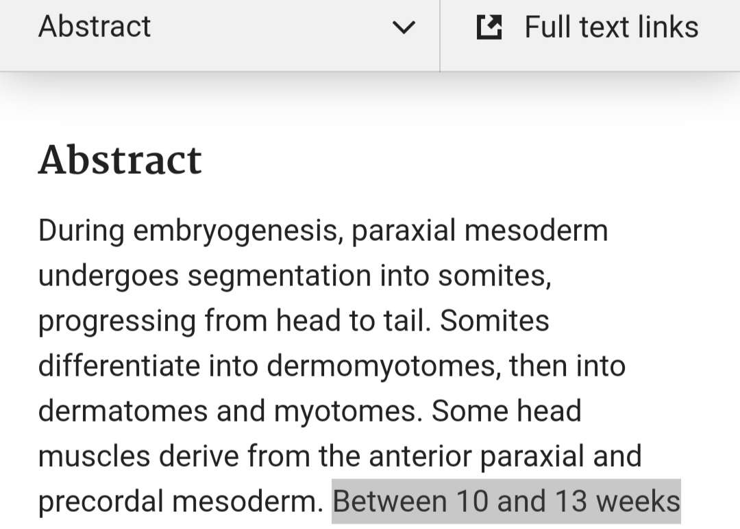
Main steps of skeletal muscle development in the human: morphologic…
During embryogenesis, paraxial mesoderm undergoes segmentation into somites, progressing from head to tail. Somites differentiate into dermomyotomes, then into dermatomes and myotomes. Some head muscles derive from the anterior paraxial and precordal mesoderm. Between 10 and 13 weeks of gestation, t …
https://www.ncbi.nlm.nih.gov/books/NBK539718/
{Embed}
https://www.ncbi.nlm.nih.gov/books/NBK539718/
Embryology, Bone Ossification
Bone ossification, or osteogenesis, is the process of bone formation. This process begins between the sixth and seventh weeks of embryonic development and continues until about age twenty-five, although this varies slightly based on the individual. There are two types of bone ossification: intramembranous and endochondral. Each of these processe…
https://www.kenhub.com/en/library/anatomy/development-of-musculoskeletal-system
{Embed}
https://www.kenhub.com/en/library/anatomy/development-of-musculoskeletal-system
Musculoskeletal system development
This article describes the embryological development of the musculoskeletal system, including the steps and processes. Learn this topic now at Kenhub!
NOTE :- the Qur’an at this verse is talking about specific type of meat which is skeletal muscle
Because Medically we classified it to Three types
1 skeletal muscle like biceps quadriceps etc
2 cardiac muscle heart
3 Smooth muscle GIT like small intestine
If you have more questions you can DM @Deleted User insha’Allah
Started a thread.