Regressive Origins of the Human Tail
Regressive Origins of the Human Tail”
🪫 “Responding to Claims of Regressive Origins of the Human Tail”
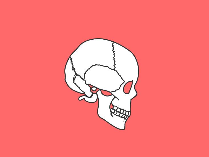
🧠 ★ One of the ridiculous pieces of evidence evolutionists use to argue that it is a remnant of a tail is the coccyx.
They claim it is useless — having once been a tail and now atrophied and without function.
Sometimes, extensions of the sacral vertebrae appear —
and they call this an “evolutionary leftover.”
But is the coccyx really useless?!
Let’s see…
- The coccyx is responsible for connecting the muscles, tendons, and ligaments in the pelvic area,
supporting and stabilizing a person while sitting.
📎 https://www.healthline.com/human-body-maps/coccyx#1
The muscles that attach to the tailbone and contribute to sitting, standing, and bowel control include:
Gluteus maximus : The gluteus maximus muscle helps maintain body alignment
Levator ani muscle : A thin muscle that helps support the pelvic organs
External anal sphincter : A flat muscle that helps with bowel function
Coccyx : A triangular muscle that supports the pelvic floor
Many of the lower pelvic muscles are attached to the coccyx, forming the “pelvic floor. ”
When these muscles don’t function properly, problems with the bowel, bladder, and sex are likely to occur.
🪫 Coccygeal malposition (subluxation) may even be a factor in pelvic floor muscle dysfunction.
Coccygeal subluxation can also directly impair bladder, bowel, and sexual function.
An important group of nerves directly in front of the coccyx is called the coccygeal ganglion ;
these nerves are partially responsible for controlling the pelvic organs.
📎 https://www.chiroweb.com/mpacms/dc_ca/article.php?id=46521
{Embed}
https://www.healthline.com/human-body-maps/coccyx
Coccyx Area, Anatomy & Function | Body Maps
The coccyx, also known as the tailbone, is a small, triangular bone resembling a shortened tail located at the bottom of the spine. It is composed of three to five coccygeal vertebrae or spinal bones.
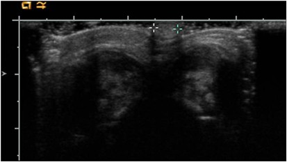
🦴 ★ Bone extensions protrude in some cases, forming a tail:
This condition is actually a spinal deformity caused by missing parts of chromosome 8 , chromosome 11 , and chromosome 17 ,
indicating a loss of information responsible for regulating spinal growth.
📎 Photograph of the chromosomal analysis showing macro deletion of the long arms of chromosome 8, 11 & 17.
📌 Sometimes it is caused by sphenoid dysplasia , which leads to skin tags.
📝 Conclusion:
The presented study indicated that true human tails are simple skin appendages without any associated spinal anomalies.
However, pseudo-tails are potentially complex lesions with a high risk of spinal dysraphisms;
warranting further diagnostic work-up and more extensive surgical technique if necessary.
🪫 The key to managing human tails is making a clear distinction between:
True tails
And pseudo-tails
📎 https://pubmed.ncbi.nlm.nih.gov/26768880/
🪫 Approximately 59 cases of human tail have been described in the medical literature.
-
49% of these were associated with spinal dysraphism
-
81% of those who underwent MRI had a tethered cord
Symptoms associated with tethered cord — including gait, genitourinary, and bowel problems —
may not appear until the third decade of life in patients with human tails and spinal dysraphism.
{Embed}
https://pubmed.ncbi.nlm.nih.gov/26768880/
The Human Tail: A Simple Skin Appendage or Cutaneous Stigma of an A…
The presented study indicated that true human tails are simple skin appendages without any associated spinal anomalies. However, pseudo-tails are potentially complex lesions with a high risk of spinal dysraphisms; warranting further diagnostic work-up and more extensive surgical technique if necessa …

Caudal appendages also occur as typical or recurrent symptoms of some syndromes.
There is no imaging or surgical evidence to suggest the frequency of tethered cord in these syndromes,
which often limits the patient’s life expectancy.
🪫 Symptoms related to cord tethering — including ambulatory, genitourinary and bowel problems —
may not occur until the third decade of life in patients with human tails and spinal dysraphism.
Caudal appendages also occur as typical or frequent manifestations of certain syndromes.
Very little imaging or surgical evidence exists about the frequency of tethered cord in these syndromes,
which often limits the life expectancy of the patient.
📎 https://www.nature.com/articles/jp200839
{Embed}
https://www.nature.com/articles/jp200839
Human tail–caudal appendage: tethered cord
Journal of Perinatology - Human tail–caudal appendage: tethered cord
🪫 These conditions are pathological conditions ,
and they actually have names, such as:
Spinal dysraphism
Spina bifida
Lipoma
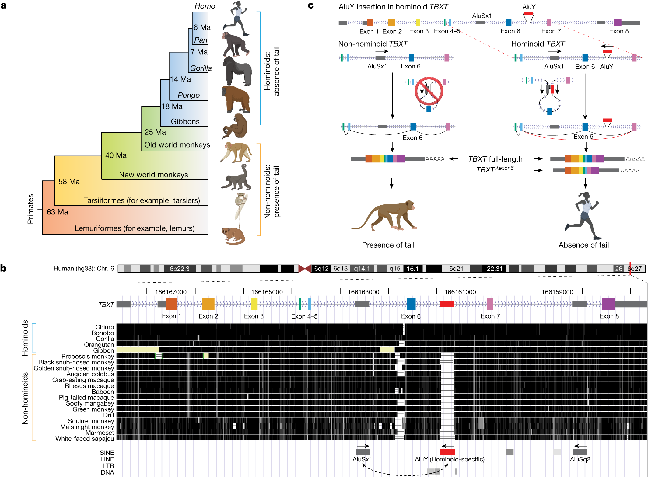
🧬 ★ They also cite this study 👇 as evidence of the evolution of the tail over time — until it was lost:
📎 https://www.nature.com/articles/s41586-024-07095-8
🪫 Before we start responding to the nonsense written in this evolutionary study (which is based on circular reasoning),
let’s first pave the way for some things to understand the research:
- Humans and mice, for example, share the vast majority of genes;
however, the same genes in humans still code for things other than those in mice, and vice versa —
even though they are the same genes —
this is what is called gene expression .
🪫 For example:
The caterpillar and the butterfly have 100% genetic similarity , with 100% genetic homology ,
yet the caterpillar is morphologically completely different from the butterfly
due to the different gene expression of the same genes.
Likewise, humans have a gene that codes for the neural tube ,
and the same gene in mice codes for the tail .
Naturally, if you alter the gene expression of a mouse to make it resemble that of a human,
the tail will be lost —
because the same gene expression in humans doesn’t create the tail, but rather the neural tube.
🪫 This is what happened in this study.
When they mimicked the expression pattern of the human TBXT gene in mice,
the tail was completely lost.
Some mice even had shortened tails,
depending on the proportion of TBXT isoforms expressed during the embryonic stage .
{Embed}
https://www.nature.com/articles/s41586-024-07095-8
On the genetic basis of tail-loss evolution in humans and apes
Nature - An insertion of an Alu element into an intron of the TBXT gene is identified as a genetic mechanism of tail-loss evolution in humans and apes, with implications for human health today.
📌 As for this statement:
“Moreover, mice expressing the exon-skipped Tbxt isoform develop neural tube defects, a condition that affects approximately 1 in 1,000 neonates in humans.”
By the way, we explained this in the introduction,
but they interpret it according to their moods to say that it is evidence of evolution…
It is natural for defects to occur in the neural tube of mice that have been imitated in gene expression similar to that found in humans.
🧠 First:
Because simultaneous changes must occur in order to avoid a deformity in the neural tube —
because every organism has its own equipped mechanism and its own genetic expressions,
and a change in one of them could lead to disasters.
🧬 Second:
The gene present in humans is only used to form the neural tube , not the tail ,
while in mice it leads to the formation of the tail .
When a defect occurs in the formation of the tail during the embryonic stages,
it is natural that problems have already occurred in the neural tube —
for example, it may lead to neural tube defects , diseases , etc.
🪫 Rather, the variation in this gene (TBXT ) is linked to the possibility of neural tube defects and chordoma ,
and not to inherit generations that do not have a tail and salvation —
but rather these generations will be disabled.
And the only logical explanation is to say that humans are created as they are —
because a change in such a gene produces generations with physical disabilities.
📎 https://www.ncbi.nlm.nih.gov/kis/ortholog/6862/?scope=7742
🪫 As for these diseases in humans, “these are diseases,”
and rare cases occur in 1 in 1,000 newborns ,
unlike what happened with these mice in the study.
{Embed}
https://www.ncbi.nlm.nih.gov/kis/ortholog/6862/?scope=7742
TBXT orthologs
Computed orthologs for TBXT - T-box transcription factor T
🎨 ★ Drawings of embryos by the German evolutionist Ernst Haeckel:
In the late 1860s, Ernst Haeckel taught the theory of “embryogenesis recapitulating evolutionary development” —
that is, during the early development of an organism,
it is supposed to retrace its evolutionary history.
He claimed that early embryos of different mammals, at one stage of development, look almost identical.
He taught that organisms go through different developmental stages in early development,
and claimed that these embryos had gill slits like fish, and tails like monkeys.
🪫 These drawings were ultimately revealed to be fake —
intended to prove the validity of evolution.
📎 https://nepalgoodnews.com/haeckels-false-embryo-drawings/
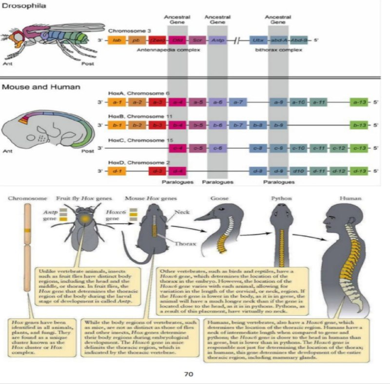
The laughing Darawneh think that the sharing of the Hox gene between organisms, and the similarity of its function, is evidence of a common ancestor!! For example, this Hox gene codes for the eye in the fly, and also codes for the eye in the mouse; beautiful… What does this have to do with a common ancestor?! The strange thing is that they ignore what is called gene expression of homologous genes, which perform different functions, such as the TBXT gene, which codes for the neural tube in humans, and codes for the tail in the mouse… They even ignore the existence of orphan genes that exist in only one type of organism, to perform functions specific to that organism only.